Estrogen is a female sex hormone that helps in the development and maintenance of the female reproductive system. There are four types of estrogens produced in the body.
This steroid hormone is produced in the ovaries, placenta, and the corpus luteum (a structure in the ovary that develops every month after the ovum is released and disappears after a few days) in premenopausal women. The liver, heart, and brain also produce minimal amounts of estrogen.
Though estrogen is most important for women, men also need it in minimal quantities to produce sperm cells and maintain sexual drive/desire.
Estrogen is essential for the growth of female secondary sexual characteristics, including:
Estradiol (E2) helps in the production of the Follicular Stimulating Hormone (FSH) and the Luteinizing Hormone (LH). Both these hormones are necessary for regular ovulation and the menstrual cycle.
The female reproductive organs have to mature healthily for a successful pregnancy. Estrogen helps in the following ways.
Mammary glands help in the production of milk after delivery. Lack of estrogen leads to immature mammary glands and problems in breastfeeding.
Estrogen helps in skeletal growth. It causes changes in the physique of girls going through puberty. Estrogen also helps maintain bone mineral density. Lack of estrogen increases the risk of osteoporosis.
Estrogen is important for the growth and functioning of axons and dendrites in the brain. Axons are nerve fibers, and dendrites are portions of nerve cells. Some studies also link estrogen to normal cognitive development, the ability to learn, remember, solve problems, think, and reason.
Estrogen reduces LDL cholesterol levels in the body and hence helps protect heart health. Estrogen also helps in maintaining blood vessel structure.
While the right amounts of estrogen is beneficial for the body, excess levels can cause harm. Excess estrogen is eliminated from the body in the three stages of detoxification. This hormone goes through different transformations in each of the stages.
The Cytochrome P450 (CYP) enzymes are responsible for the first stage of estrogen transformation. The CYP enzymes are substances that play a role in the detoxification and clearance of drugs, hormones, and other substances from the body.
Three main CYP enzymes act on estrogen. They are:
In phase 1, a hydroxyl group (-OH) is attached to the estrogen hormones, and as a result, the following intermediates are produced.
These intermediates are called metabolites. These metabolites are very reactive and need to be quickly cleared by phase 2 and phase 3 detoxification. When produced in excess, these metabolites react with the DNA and lead to an increased risk of cancers.
In Phase 2 detoxification, these hydroxyl metabolites become less reactive and more stable with the addition of a methyl group. The Catechol-O- MethylTransferase (COMT) enzyme is responsible for the methyl group addition.
The substances produced as a result are:
Unlike the earlier metabolites, these are safer and neither react with the DNA nor lead to abnormal DNA transformations.
Apart from methylation, estrogen also goes through sulphation using the sulfotransferase (SULT) enzymes. The SULTs attach estrogen with sulfate molecules. Conjugation is the process of making the hormone more water-soluble to make it easy for excretion. Estrogen sulphation creates estradiol, and this is sent back to circulation in the body.
Estrogen also goes through a third process called glucuronidation in phase 2. In this process, estrogen conjugates with glucuronic acid using the UDP-glucuronosyltransferase (UGT) enzyme. This makes it easier for estrogen to be eliminated from the body.
However, another enzyme called β-glucuronidase that is found in the breast glands and the gut can reconvert the conjugated estrogen back into free estrogens. This reconversion leads to abnormally high estrogen levels in the body.
Phase 3 is the final stage of detoxification. Here, estrogen metabolites leave the body through urine or bile. The antiporter proteins help in phase 3. These are also called exchanger proteins. These proteins carry substances in and out of the cells and help eliminate the final estrogen metabolites.
The β-glucuronidase enzyme found in the breast cells and gut can create problems in the functioning of the antiporter proteins. As a result, it leads to excess estrogen circulation.
Changes in genes like CYP1A1, CYP1B1, CYP3A4, COMT, SULTs, and UGTs can lead to estrogen production problems and increase or decrease the hormone levels in the body.
Low levels of estrogen may result in:
High levels of estrogen may result in:
According to the National Cancer Institute, there will be 281,550 new cases of breast cancer diagnosed in the United States in 2021. Latest studies report that estrogen levels in the body may play a role in breast cancer.
While age and genetics play the most important role in causing breast cancer, lifetime exposure to estrogen is also an important cause.
The CYP enzymes convert estrone to 2-hydroxyestrone, 4-hydroxyestrone, 16-hydroxyestrone, or estriol. They also convert estradiol to 2-hydroxy estradiol and 4-hydroxy estradiol. This conversion process releases free radicals that increase the risk of all types of cancers, including breast cancer.
Estrogen Receptor α (ERα) is a group of proteins that estrogen activates. Once activated, ERα attaches itself to the DNA and controls the activity of different genes.
According to studies, certain changes in the estrogen hormone can lead to changes in the ERα too. The ERα causes errors in DNA replication and leads to multiple, abnormal divisions of cells. This leads to the formation of tumors.
Breast cancer that develops because of estrogen receptors is called estrogen receptor-positive breast cancer (ER-positive breast cancer). This accounts for 80% of all breast cancers.
An estrogen levels test will measure the estrogen levels in the blood and urine. This test can individually measure E1, E2, and E3 levels. Getting the test will tell you if you are at a higher risk for estrogen-associated health conditions.
Specific genetic changes can affect the levels of estrogen levels in the body. Some people will have naturally higher estrogen levels that could increase the risk of breast and other types of cancers. Genetic testing will help confirm if the person is at higher risk for abnormal estrogen metabolism.
A 2019 meta-study analyzed the relationship between diet and risk of breast cancer. It included results from 32 articles and reported the following:
Dietary phytoestrogens are types of foods that cause estrogen-like effects in the body. Plant-based foods are the primary sources of dietary phytoestrogens. Studies report that these phytoestrogens help balance the endocrine system and bring down the risk of estrogen receptor-positive breast cancer.
Other studies mention that dietary phytoestrogens affect the 2-hydroxy estrone to 16α-hydroxy estrone ratio. This regulates estrogen metabolism and reduces the risk of breast cancer.
A small study picked up 272 participants, including 37 semi-vegetarians (women who consumed less than 30 g of meat a day) and 235 non-vegetarians (whose primary diet included red meat every day). This study reported that serum estrone and estradiol levels in semi-vegetarians were lower when compared to non-vegetarians.
Another study divided 115 women into two groups - control and intervention. Women in the intervention group were put on a Mediterranean diet (including fish, fresh fruits, vegetables, greens, and dairy). The control group followed a regular western diet style. At the end of 6 months, women in the intervention group had up to 40% lower estrogen levels than the control group.
Foods rich in fiber help with better estrogen metabolism and bring down excessive estrogen levels in the body. Fiber can flush out cholesterol deposits (cholesterol leads to estrogen production). Some plant-based fiber foods that can aid estrogen metabolism are:
When we are born, we inherit (receive) the genes from our parents, but we don't inherit their age.
In other words, how do older parents give rise to young offspring?
Scientists have tried to explain this by a process called "natural rejuvenation event." This event sets our biological age to zero, which is the beginning of aging in mammals.
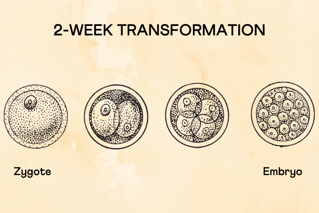
This event happens as the zygote develops to form an embryo. A zygote is a single-celled organism resulting from a fertilized egg. The zygote goes through a cellular multiplication process to give rise to the multi-celled embryo.
Researchers are studying the possible application of this "age reversal event" as a therapeutic intervention for age-related diseases, such as arthritis or Parkinson’s.
Aging is something we all experience but actually know very little about. Hair greying, wrinkles formation, joint changes, etc., are signs of aging. Aging, on the other hand, encompasses all the processes that occur in the body that result in these signs - it is a combination of physiological changes in our bodies and environmental factors.
Aging eventually leads to physical denaturation, dysfunction, and ultimately mortality (death).
As we age, every cell in our body gets old due to the progressive accumulation of damage through the intervening years. But this cellular damage does not get passed on to human offspring to the next generation – the absence of this cellular damage in human offspring has baffled scientists for generations.
That's because germline cells (sex cells - egg and sperm that form the embryo/zygote) rejuvenate in the offspring after conception.
During the initial stages of embryogenesis (the formation of the embryo), life hits a reset button — the rejuvenation event in the embryo. When an embryo is attached to the uterus, the rejuvenation event sets the developing embryo to grow at its youngest biological age – zero. This rejuvenation of the embryo marks the beginning of the mammalian age.
The epigenetic aging clock is a biochemical test used to measure age. It was developed with conserved cytosines - units in DNA whose methylation levels change with age. Experts use these epigenetic clocks to predict the approximate ages of mouse embryos at their early stages of development.
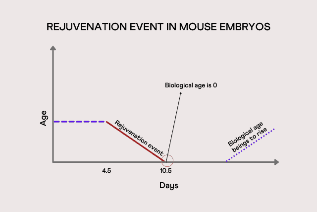
They found that the age of mouse embryos stayed constant for the first few days after fertilization. However, by around 6.5 to 7.5 days into development, the biological age of the embryos witnessed a dip, indicating that they are undergoing some type of rejuvenation event.
The researchers say that the biological age of mice is set to zero, somewhere between 4.5 to 10.5 days after fertilization.
Eventually, during development, the biological age begins to slowly rise. However, the point exactly at which this growth happens is still unclear.
At present, similar data for humans is unavailable as studying human embryos at such early stages is prohibited. Recent studies have shown that the epigenetic clocks in cord blood can be used to estimate gestational age at birth.
When embryos can reset the biological clock, there is a hope to reverse the errors that occur due to cell damage, restoring the cells back to their agile state, eliminating signs of aging.
This hypothesis could possibly help researchers develop treatments for age-related diseases, such as arthritis or Parkinson’s - A paradigm shift in treating age-related illnesses.

Triglycerides are types of fat that are commonly found in the human body. The name ‘triglyceride’ means a combination of three kinds of fats combined with a form of glucose called glycerol. The three kinds of fats are - unsaturated fats, saturated fats, and a combination of both.
Triglycerides are majorly present in the fat deposits in the body. These are also present in the blood. These hold on to unused calories in the body and reserve them for future use.
There are two ways your body receives triglycerides.
Most of the foods we eat are sources of triglycerides. Excess fat in food directly gets stored as triglycerides, while excess carbohydrates and sugars are converted to triglycerides by the liver and stored.
Your triglyceride levels increase when you consume more calories than what your body can burn. When you exercise, you burn extra calories and hence prevent the increase in triglyceride levels.
A 1982 study analyzed the levels of triglycerides in endurance athletes after long sessions of working out. The study concluded that there was a significant decrease in serum triglyceride levels after 1-hour and 2-hour sessions of exercise.
Another study considered the effects of aerobic exercise on serum triglyceride concentration levels. The study included 38 patients with existing Coronary Heart Disease (CHD). One group underwent aerobic training for eight weeks, and the other group remained sedentary.
The study concluded that people who exercised showed a lowered concentration of triglyceride levels.
A large-scale 2014 study analyzed the results of 13 independent studies relating aerobic exercise, resistance training, and combined exercise on triglyceride levels. According to the study:
Triglycerides are major energy sources in the body. Every unit of triglyceride contains more energy than one unit of protein or carbohydrates. That is why you feel full and sated when you eat a fat-based meal.
When you consume triglycerides, they reach the intestine. Here, they are combined with particles called lipoproteins. Lipoproteins transport lipid (fat) molecules through the plasma to other parts of the body.
Lipoproteins take the triglyceride particles to different muscles and tissues that need energy.
Triglycerides are stored in the fat tissues and the liver in the body. If you are suddenly deprived of food and are starving, stored triglycerides are broken down in the fat tissues and are used for energy. Triglycerides are hence very important backup energy sources.
According to the National Cholesterol Education Program (NCED), here are the different categories based on recommended triglyceride levels.
Familial Hypertriglyceridemia - This is an inherited condition where the liver overproduces Very-Low-Density Lipoproteins (VLDL). VLDLs are responsible for carrying triglycerides to the tissues of the body from the liver. High VLDL levels also increase blood triglyceride values.
The Cysteine and tyrosine-rich 1 gene (CYYR gene) contains instructions for the production of the CYYR protein. The exact functionality of this protein is not understood yet.
rs222158
The A allele of the SNP rs222158 of this gene affects triglyceride training-response. This allele is associated with decreased triglyceride levels in response to exercise.
The GLT8D2 gene (Glycosyltransferase 8 Domain Containing 2 gene) is responsible for the production of the GLT8D2 protein. This protein plays a role in glycosyl transfer.
rs2722171
The RBFOX1 gene (RNA Binding Fox-1 Homolog 1 gene) produces the RNA binding protein fox-1 homolog 1. Abnormalities in the protein can lead to neurodegenerative diseases.
rs1906058
The type of exercise - If you want to bring down your triglyceride levels with exercise, choosing the right workout regime is important.
Aerobic exercises are the best choices for lowering triglycerides. You can also try resistance exercises. High-intensity exercises are better as they quickly burn fat and help lower your triglyceride levels.
Excess fat consumption - When you keep consuming excess fatty-foods, even when you exercise rigorously, the body will always have excess fat reserves, and hence the triglyceride values will not decrease.
Excess carbohydrate consumption - People who consume excess carbohydrates and simple sugars are at high risk for developing high levels of triglycerides. This condition is called carbohydrate-induced hypertriglyceridemia.
Studies show that when more than 55% of the energy consumed is through carbohydrates, the body works in converting excess carbohydrates into fat.
As a result, even if you are controlling the amount of fat you consume and are working out, your triglyceride levels will not reduce as much as you expected.
Smoking - A study compared the fasting triglyceride levels in smokers and non-smokers. It concluded that smokers had high fasting triglyceride levels when compared to non-smokers.
Another study analyzed the effects of smoking on aerobic capacity and concluded that the muscles in the bodies of smokers receive less oxygen than in non-smokers, and hence smokers are unable to perform intensive workouts.
Smoking increases triglyceride levels and brings down a person’s ability to exercise effectively. As a result, in smokers, exercising does not cause a considerable reduction in triglyceride levels when compared to non-smokers.
Triglyceride levels can be identified with a simple blood test. When your blood shows higher levels of triglycerides, here are risk factors to consider:
Try a combination of exercise and a calorie-restricted diet
The more regularly you work out, the more fat your body will burn. Studies show that when you don’t exercise and go on a calorie-restricted diet, it doesn’t affect triglyceride levels as much as exercise does. A combination of moderate to high-intensity exercise and a calorie-restricted diet plan works wonders.
Change your diet plan
Opt for a high protein and moderate fat and carbohydrate diet. A high-fiber diet is also considered beneficial. Restrict consuming trans and saturated fats. These changes help you exercise better and, as a result, reduce your triglyceride levels.
Slowly build your stamina
Sometimes, existing health conditions, age, and other related factors can prevent a person from taking up exercising. In that case, slowly build up your stamina. Start with low-intensity workouts like walking and then move on to aerobic and resistance training. With time, you will be able to work out enough to lower your triglyceride levels.
https://en.wikipedia.org/wiki/Triglyceride
https://www.heartuk.org.uk/cholesterol/triglycerides
https://www.uofmhealth.org/health-library/zp3387
https://www.mayoclinic.org/diseases-conditions/high-blood-cholesterol/in-depth/triglycerides/art-20048186
https://www.urmc.rochester.edu/encyclopedia/content.aspx?contenttypeid=56&contentid=2967
https://www.medicinenet.com/how_to_lower_triglycerides_naturally/article.htm
Detoxification is a natural process that happens in the human body to get rid of harmful substances known as toxins. The buildup of toxins can lead to an imbalance in the body, giving rise to diseases. Our bodies have the capacity to rid of these harmful substances through detoxification pathways. Detoxification has a significant impact on human health.
We get exposed to chemicals via the air we breathe, medications we take, cleaning products we use, personal care products we put on our skin, and several other products we are exposed to on a daily basis.
There are two types of toxins in the body: Exogenous and Endogenous.
Exogenous Toxins: These are absorbed by the body from external sources like water, air, food, or direct contact. Drugs, chemicals, and microorganisms are examples of exogenous toxins.
Endogenous Toxins: These toxins are produced in the body as by-products of metabolism. Endogenous toxins also include the metabolic by-products of the bacteria that live in our bodies.
Toxins are naturally fat-soluble and, therefore, cannot be eliminated from the body easily. Due to this characteristic, toxins have an affinity for the fat cells in the body and can be stored in them for years.
Toxin buildup in the body causes liver disorders, obesity, irregular sleep patterns, fatigue, and cancer. Detoxification helps eliminate toxins by converting them into water-soluble substances.
The usual sequence of events in the detoxification process is as follows:
The detoxification process can be grouped under three important phases – Bioactivation, Conjugation, and Antiporter Activity.
In phase 1 detoxification, the toxins are prepped for phase 2. This phase of detoxification is performed primarily by the Cytochrome P450 (CYP) enzymes.
The side-effect of this stage is the formation of free radicals, which are reactive molecules that can harm the body.
Conjugation occurs in six stages. In this phase, the toxins are deactivated and made more water-soluble to facilitate easy excretion via bile (small intestine) or via urine (kidneys). This phase of detoxification is governed by various enzymes, including the glutathione transferases and UDP-Glucuronosyltransferases(UGT). Some genes that play an active role in the conjugation phase include SULT1A1, n-acetyltransferase genes, and COMT.
Phase 3 of detoxification is the excretion phase that takes place in the liver, kidneys, and intestines. During this phase, the water-soluble toxins are transported across membranes of the cells in the liver, the gastrointestinal tract, the kidneys, and the blood-brain barrier. This phase of detoxification is governed by the ATP-binding Cassette protein (ABC) and the Solute Carrier (SLC10) gene families.
The ABC family of genes is primarily involved in influencing the absorption and outflow of the processed toxins from the cells. Two genes from the SLC family are involved in the production, absorption, and excretion of bile salts (the main pathway for excretion of toxins) out of the body via the small intestine.
The inability of the body to detoxify efficiently is termed impaired detoxification. Studies have linked impaired detoxification to health conditions like certain types of cancer, autism, Parkinson's, fibromyalgia, and immune dysfunction syndrome.
The NFE2L2 gene regulates the activity of the NRF2 gene that produces the NRF2 protein. This protein plays an important role in the body's detoxification process. The NRF2 gene activates four other genes that produce proteins required in the detoxification process.
Some abnormal changes in the NFE2L2 gene reduce the expression and activity of the NRF2 gene. Reduced NRF2 activity impairs the body's ability to detox and defend itself from oxidative stress.
There are many ways by which you can boost your body's detoxification process. Some of the most effective ones include:
Use our free Gene Tool to check your detox gene variants.

Psoriasis is a global health problem prevalent among people of all ages, affecting 100 million individuals worldwide. Previous studies have suggested that a high-fat and high-sugar diet can lead to disruption in the gut's microbial community, contributing to inflammatory skin diseases such as psoriasis. Recent research shows that switching to a balanced diet can restore gut health and reduce inflammatory symptoms.
Psoriasis is a chronic skin disease that causes the skin cells to multiply faster than normal. This leads to an overgrowth of skin cells that leads to scaling on the skin's surface. The red, itchy, scaly patches are usually found on the elbows, scalp, knees, and lower back. Abnormalities in the immune system, emotional stress, blood pressure medications, hydroxychloroquine, and strep infections may trigger psoriasis.
Over time, 10-20 percent of affected individuals develop psoriatic arthritis, an inflammatory type of arthritis. It is associated with symptoms similar to arthritis-like joint pain, stiffness, swelling, etc.
Western diets include high intakes of red meats, processed foods, fatty foods, refined grains, potatoes, high sugar drinks, and low intakes of vegetables, fruits, nuts, fish, and whole grains. On the other hand, a balanced diet is a healthier option. It includes fresh vegetables and fruits, legumes, whole grains, lean proteins, and nuts.

Previous studies show that a western diet characterized by high sugar and fat content leads to significant skin inflammation and psoriasis.
Food is a major regulating factor of the microorganisms living in our intestines. Consuming a western diet can cause rapid change to this microbial community and its functions. This disruption in microbial balance contributes to gut inflammation. It also affects skin immunity, leading to skin inflammation.
A study led by Dr. Hwang and his team of researchers at UC Davis Health examined if switching to a balanced diet can restore the gut microflora and help reduce skin and joint inflammation.
The team used mouse models to study the effect of diet on psoriasis and psoriatic arthritis. The researchers subjected the mice to the following:
The researchers then randomly divided the mice into two groups; one group continued the western diet for another four weeks, and the other group switched to a balanced diet for the same period.
The following were observed at the end of 10 weeks:
Coenzyme Q10 also called Ubiquinol is a natural antioxidant found in all cells of the body. It plays an important role in metabolism. It is a coenzyme, meaning it helps enzymes work more effectively.
CoQ10 is stored in the powerhouse of the cell, the mitochondria. Mitochondria are responsible for producing energy. This coenzyme plays a major role in mitochondrial bioenergetics. It helps transport electrons in the mitochondria to facilitate the process of producing ATP, which is the main source of energy for cells.
As an antioxidant, CoQ10 protects the cells from oxidative damage by neutralizing free radicals. Oxidative damage can lead to inflammation, tissue damage, and cell death.
CoQ10 levels are higher in the brain, heart, kidneys, muscle, and liver. These are the organs that have many mitochondria as they need a lot of energy for functioning.
CoQ10 is found in two forms, ubiquinol, and ubiquinone. Ubiquinol is the active form and performs various functions like reducing oxidative damage. Ubiquinone is the oxidized version.
CoQ10 is naturally produced in the body. The levels of CoQ10 decrease with age. Several disease conditions also interfere with the levels of CoQ10. In these cases, people usually take CoQ10 in the form of supplements.
People with CoQ10 deficiency experience physical fatigue and muscle weakness, even while doing less intense physical activities like walking. It can also lead to mental fatigue, affect your memory and ability to concentrate.
CoQ10 deficiency limits the body’s ability to fight against oxidative damage and this may lead to faster progression of health conditions linked to oxidative stress.
Lower levels of CoQ10 have been linked to nutrient deficiencies, heart diseases in patients who take statin drugs to lower their cholesterol levels, and other disease conditions.
Research shows that CoQ10 supplementation can improve power, recovery after exercise, reduce oxidative damage, and increase energy.
Training can lead to oxidative stress in the muscles. CoQ10, being an antioxidant, can decrease oxidative stress in cells and improve mitochondrial function to provide energy and fuel your workout.
Studies show that CoQ10 can increase exercise performance by reducing fatigue and increasing power.
Mutations in genes involved in CoQ10 production are found to interfere with levels of CoQ10.
The NQO1 gene encodes for the NAPDH quinone dehydrogenase 1 protein. This protein is involved in the reduction of Coenzyme Q10. Variations in this gene affect levels of CoQ10.
The CoQ10 levels of TC carriers were found to be significantly lower than CC carriers.
| Genotype | Implication |
| CC | |
| TC | Lower CoQ10 levels than CC genotype |
| TT | Lower levels of CoQ10 |
Age: Production of CoQ10 naturally in the body decreases with age.
Health conditions: Certain nutritional deficiencies, heart disease, brain disorders, cancer, diabetes are associated with low levels of CoQ10.
Statin medications: Statin is commonly used to treat high cholesterol in people with heart diseases. Statins block the production of CoQ10 and can lead to CoQ10 deficiency.
CoQ10 is naturally produced in the body. People with certain disease conditions take supplements to help with treatment. Athletes generally take CoQ10 supplements to improve their performance. CoQ10 can be supplemented through diet or medication.
Food sources of CoQ10 include
There are various CoQ10 supplements available in the market in the form of tablets. CoQ10 supplements are generally safe and have minimal side effects.
Make sure to talk to a healthcare professional about the correct dosage and possible side effects of taking these supplements. CoQ10 supplements may interfere with the effectiveness of certain other drugs like blood thinners, blood pressure medications, and thyroid medication that are used to treat health conditions.
https://www.nccih.nih.gov/health/coenzyme-q10
https://www.healthline.com/nutrition/coenzyme-q10#TOC_TITLE_HDR_1
https://www.webmd.com/diet/supplement-guide-coenzymeq10-coq10#1
https://www.mayoclinic.org/drugs-supplements-coenzyme-q10/art-20362602